Published in Journal of Vascular and Interventional Radiology in May 2015
- Ivan Kuang Hsin Huang, MBBS, MMed, FRCR
- Mahendran Nadarajah, MBBS, FRCR
- Li Tserng Teo, MBBS, FRCS
- Dokev Basheer Ahmed Aneez Ahmed, MBBS, FRCS, FAMS
- Uei Pua, MBBS, MMed, FRCR, FAMS
- Department of Diagnostic Radiology (I.K.H.H., U.P.);
- Trauma and Acute Care Surgery Service, Department of General Surgery (L.T.T.); and Thoracic Surgery and Thoracic
- Oncology Service, Department of General Surgery (D.B.A.A.A.)
Editor:
We report a case of an acute large juxtacardiac right inferior pulmonary vein pseudoaneurysm following blunt chest trauma that was successfully treated with direct percutaneous coil embolization. Our institution does not require ethics approval for case reports such as this.
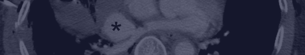
A 49-year-old man sustained blunt chest wall injury during a motor vehicle accident and presented with a flail chest. He was subsequently intubated at the emergency department because of an inability to maintain oxygen saturation despite supplementary oxygen. Contrast-enhanced computed tomography (CT) of the thorax revealed a 3.21-cm 2.6-cm wide-necked juxtacardiac right inferior pulmonary vein pseudoaneurysm (Fig 1), together with multiple rib and sternal fractures, extensive pulmonary contusion, right hemothorax, left hydropneumothorax, and hemopericardium. After multidisciplinary consultation among trauma and cardiothoracic surgeons and interventional radiologists, it was agreed that surgical repair in the acute setting posed a high risk as a result of the central location of the lesion, current unstable hemodynamic status, and likely extensive adhesions from a previous pleurodesis. The consensus was to attempt embolization to stabilize the patient’s condition for eventual surgical repair.
Direct percutaneous access followed by coil embolization was chosen after considering available resources andexpertise limitations for various endovascular approaches. For pulmonary venous access, the needle trajectory was planned by correlating internal landmarks on cone-beam CT with the thoracic CT images; the pseudoaneurysm was midway between the bifurcation of the right main bronchus and T6 vertebral body and was defined as the target on the navigation software.
Download/ View Journal Article (PDF, 1.67 MB)
References
- Guyader, A., Bertin, F., Laskar, M. et al, Blunt chest trauma: a right pulmonary vein rupture. Eur J of Cardiothor Surg. 2001; (1054-6 B).
- McKeown, P., Rosemurgy, A., Conant, P. Blunt traumatic rupture of pulmonary vein, left atrium and bronchus. Ann Thorac Surg. 1991;52:1171–1172.
- Brockenbrough, E.G., Braunwald., E. A new technic for left ventricular angiocardiography and transseptal left heart catheterization. The Am J of Cardiol. 1960;6:1062–1064.