What is an Exercise Stress Myocardial Perfusion Imaging Test?
The exercise stress myocardial perfusion imaging test assesses the blood flow to the heart muscle when the heart is made to work harder ('stressed'). This test determines the likelihood of a blockage in one or more of the coronary arteries. Blood flow in the heart is assessed by injecting a very small amount of a radioactive chemical, usually technetium or thallum, which is absorbed by the heart. The heart is then imaged using a special gamma camera.
Patients usually undergo a treadmill exercise to create stress to the heart and to assess the adequacy of blood supply to the heart muscle.
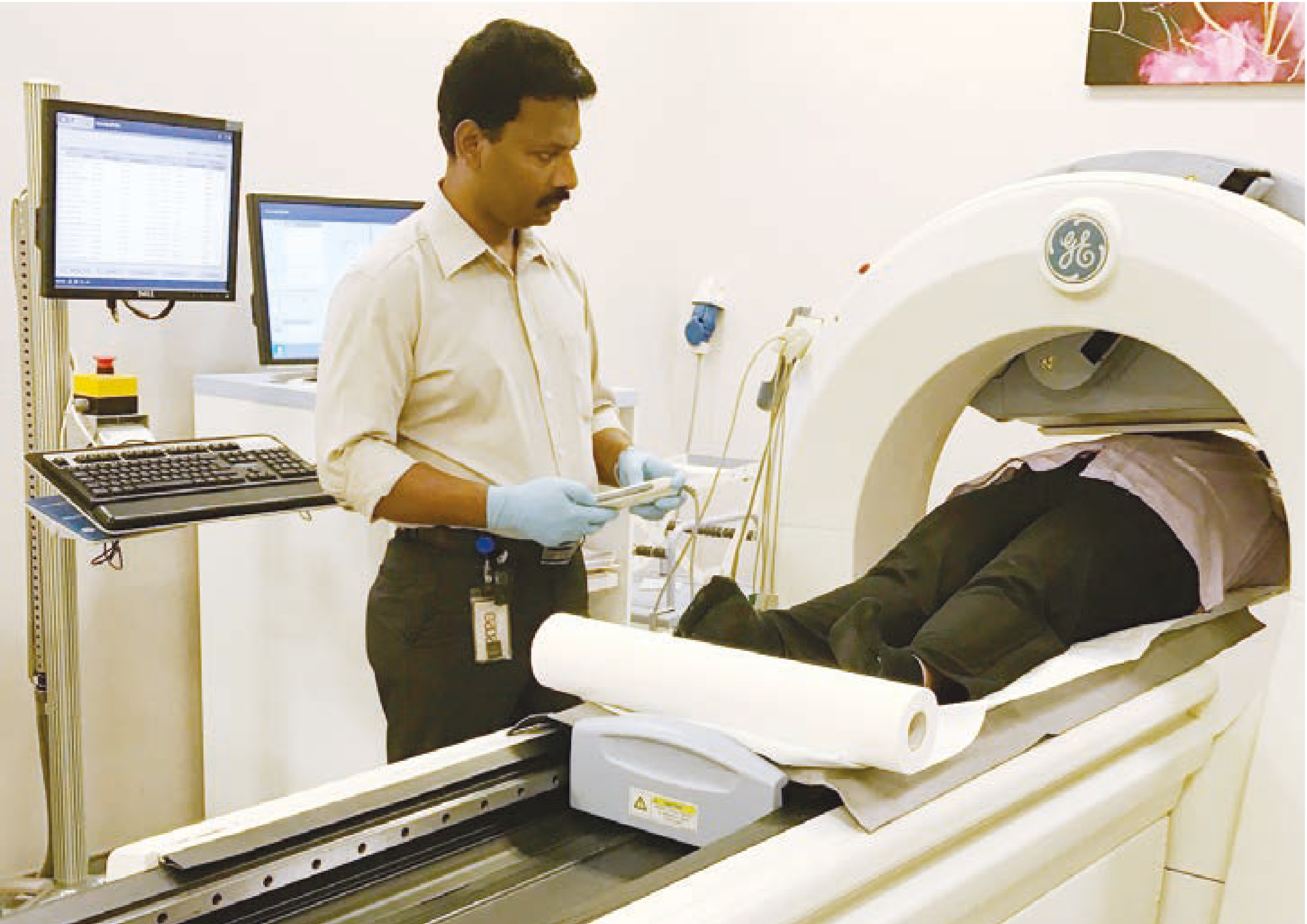
Imaging table and the gamma camera used to capture
images of the heart.
What is the Purpose of the Test?
Coronary arteries are blood vessels supplying the heart. They can be abnormally narrowed in a medical condition called atherosclerosis. For patients with atherosclerosis, there may not be enough blood flow to the heart during physical exertion, leading to symptoms of chest discomfort and shortness of breath.
This test assesses the blood flow to the heart and also gives the doctor an indication of how strongly the heart is beating (the ejection fraction). These are the two most important pieces of information a doctor needs when evaluating a patient for heart disease.
What can I Expect?
The test is performed in the Nuclear Cardiology Laboratory. It consists of the stress phase and the rest phase, both of which are usually completed on the same day. However, in some cases due to technical factors, it may be necessary to perform each phase on separate days.
Before the Test
Your height and weight will be taken. A small plastic tube or cannula is inserted in one of the veins on your hand for injection of the radioactive chemical.
To facilitate the attachment of electrocardiogram (ECG) electrodes to the chest, male patients will be asked to remove their shirts. Female patients may be asked to change into special gowns for the purpose of attaching electrodes. Once your blood pressure and baseline ECG is recorded, you will then proceed with either the resting or stress phase of the test.
During the Test
1. Resting Phase
The radioactive chemical will be injected into the vein through the cannula and shortly thereafter, you will be asked to lie on the imaging table of the gamma camera for about 15 minutes. It is important to lie as still as possible for good images to be captured. After a 2 hour interval, you may proceed to the 'stress' phase of the test.
2. Stress Phase
You will be asked to walk on a treadmill, the speed and incline of which will increase after every 3 minutes. Throughout the test, your ECG and blood pressure will be monitored and a doctor will be present during this phase of the test. You must inform the doctor supervising the test if you experience any symptoms. At the peak of the exercise, the doctor will inject the radioactive chemical into your vein. For the radioactive chemical to function, you must continue to exercise on the treadmill for one to two minutes after the injection.
After the exercise, you will continue to be monitored for a few minutes during the recovery period. After resting for 30 to 45 minutes, images of your heart will be taken again with the gamma camera. You may consume some water during this period.
Taking images of the heart while facing downwards on the imaging table may be needed to improve accuracy of the test.
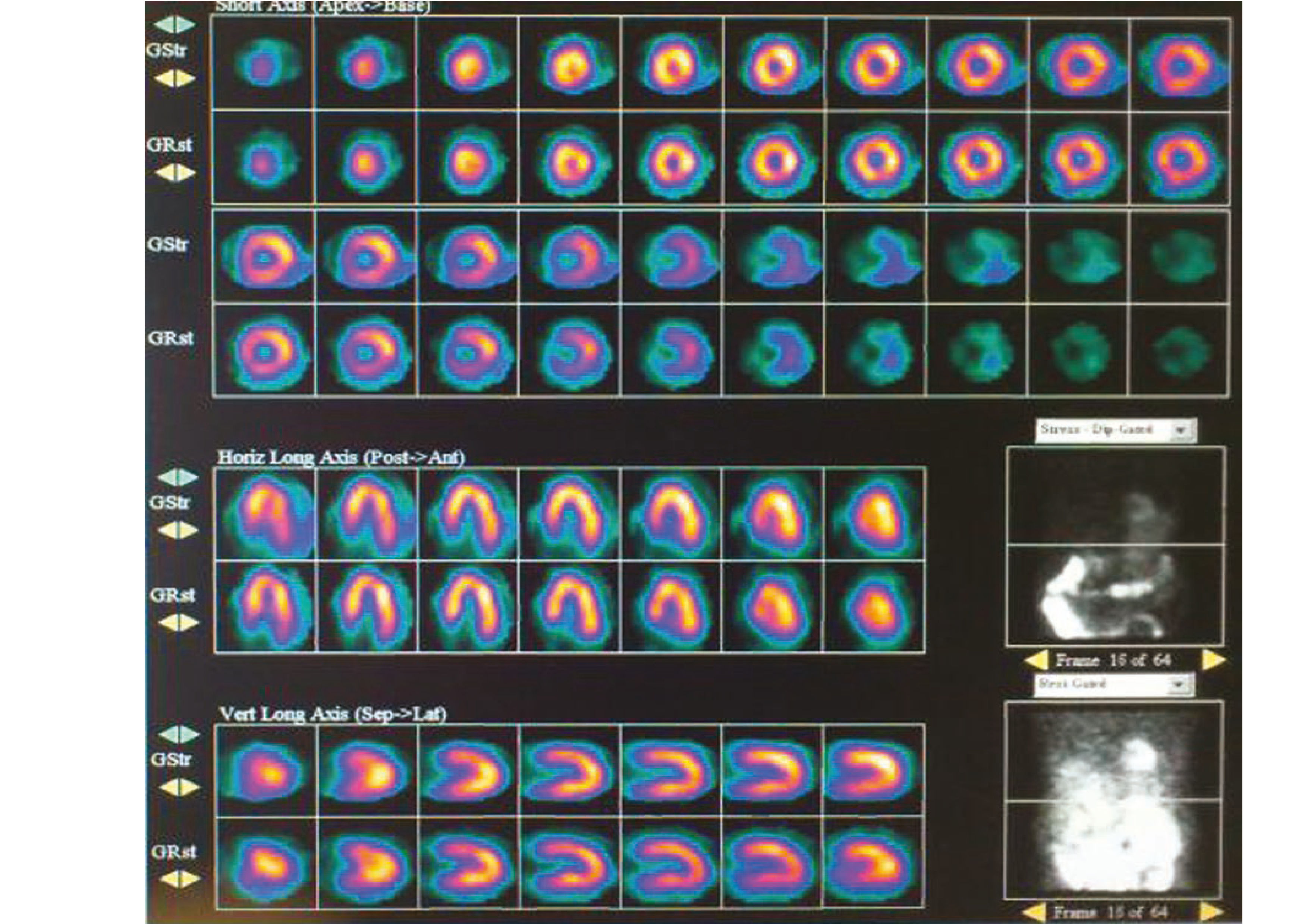
Cross-sectional views of heart muscle showing normal
blood flow and normal perfusion imaging.
Inform Your Doctor On:
- Any medications that you are taking for high blood pressure and/or heart disease, which may slow down the heart rate. These medications should be omitted 24 to 48 hours prior to the test, except in specific clinical situations. Please check with your doctor regarding this.
- Your conditions that can make exercise difficult, such as joint and leg circulation issues or problems with balance. Also, the exercise test should not be performed if you have a fever, a virus and other accompanying acute illnesses. Please check with your doctor regarding this.
- Your pregnancy. You should not undergo this test if you are pregnant.
Preparing for an Exercise Stress Myocardial Perfusion Imaging Test
- Come in sports attire and running shoes.
- You can have a light meal before the test. Avoid taking food or drinks which contain caffeine (Coffee, Tea, Chocolate, Coke) for at least 12 hours before the test. If you smoke, avoid smoking for at least 6 hours before the test.
What are the Potential Risks/ Complications?
Exercise testing is generally very safe. The risk of a major complication, including a heart attack, is very low. Experienced medical staff present during the test will identify any complications at an early stage and hence terminate the test, if needed.
The radioactive chemical given is safe and has no known immediate side effects. Exposure to radioactivity, theoretically, has a small risk of causing cancer. The risk is extremely small and, for appropriately selected patients, the potential benefit of diagnosing and then treating your heart condition far outweighs this small risk.
When Will I Know the Results?
Your results and the next step of your treatment will be discussed at the next appointment with your doctor. Preliminary results will not be given on the day of your test. However, in the event of a very severe abnormality requiring urgent medical attention, appropriate measures will be taken. These measures include either prompt admission to the hospital or rescheduling your appointment with your doctor to an earlier date.