What is Retinal Vein Occlusion?
Occlusion (blockage) of a retinal vein is one of the most common causes of sudden painless reduction of vision in the older population. It is the second most common cause of blindness from retinal vascular disease after diabetic retinopathy.
In
Retinal Vein Occlusion (RVO), a blockage, commonly a clot, forms in the retinal vein.
This disease can affect a very important structure in the eye: the
retina. The retina is the thin membrane that lines the inner surface of the back of the eye. It is akin to the film in a camera that
receives light rays and transmits the signals to the brain thus allowing one to see.
Blockage of one of the veins draining blood from the retina causes bleeding and swelling of the retina. This can cause slow or sudden loss of vision.
It can occur at any age, though typically in the older age group (usually above 60s).
What Causes Retinal Vein Occlusion?
There are several factors that can cause retinal vein occlusion. Common ones are hypertension, diabetes and glaucoma. Rarer causes include infections and blood conditions where clotting occurs easily.
What are the Types of Retinal Vein Occlusion?
-
Central Retinal Vein Occlusion (CRVO)
The central retinal vein, which is formed from the four branches that drain blood from the retina, is obstructed.
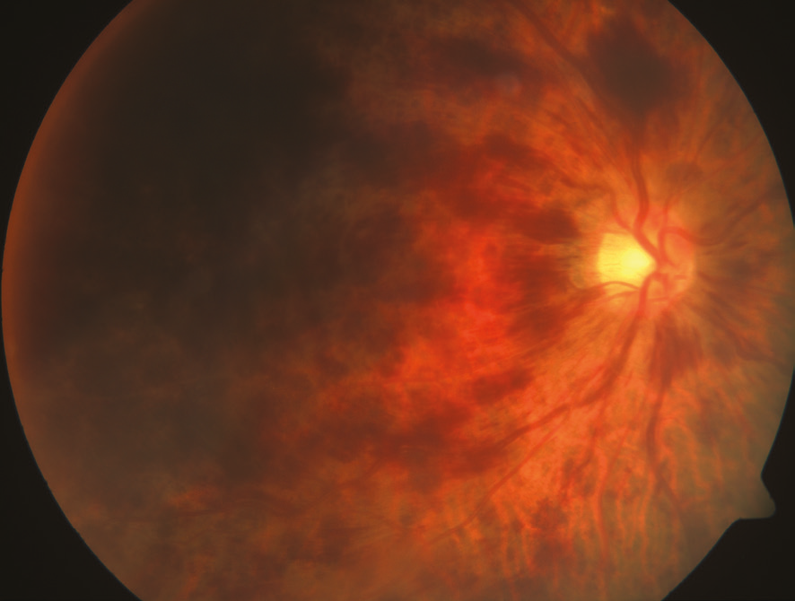
Central Reinal Vein Occlusion in the right eye.
-
Branch Retinal Vein Occlusion (BRVO)
One of the four retinal veins that draw blood from the retina is blocked. This is the most common form and it is painless.
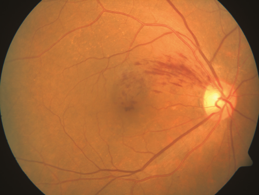
Branch Reinal Vein Occlusion in the right eye.
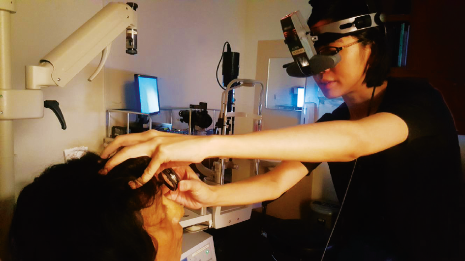
Diagnosing Retinal Vein Occlusion
Your eye doctor may be the only person who can tell you if you have a retinal vein occlusion. He or she can do so in the following ways:
- Checking your vision.
- Examining the appearance of your retina.
- Using retinal imaging to identify retinal ischaemia (inadequate blood supply) and leaks from the vein occlusion.
- Using high resolution imaging to measure the retinal swelling.
How Does One Lose Vision from Retinal Vein Occlusion?
-
Macular oedema. The macula is the most sensitive central area of the retina important for sharp vision. Blood and fluid leaking in the macula causes swelling, a condition called macula oedema, that causes blurring or loss of vision.
-
Neovascularisation. RVO can cause the retina to develop new, abnormal blood vessels, a condition called neovascularisation. These new vessels are fragile and burst easily, leaking blood into the vitreous. With severe neovascularisation, the retina can detach from the back of the eye.
-
Neovascular glaucoma. New blood vessels in the front of the eye can cause pain and a dangerous increase in pressure inside the eye.
The complications of RVO, especially if not treated, can lead to irreversible loss of vision.
That is why it is so important to continue with the eye doctor’s follow-up when you are diagnosed with RVO.
How Will My Eye Doctor Treat My Retinal Vein Occlusion?
Unfortunately, there is no way to unblock retinal veins.
Once diagnosed, a patient requires strict
follow-up with his eye doctor. It is important to identify the cause and treat any health problems that may be related to the RVO. This aims to
reduce the risk of recurrences to the affected or the other eye.
Treatment is offered for the complications. Some of the treatments for retinal vein occlusion include laser therapy and intravitreal injections of medications.
With appropriate treatment, there can be a better chance of reducing visual loss and complications from the condition.
How Can I Prevent Retinal Vein Occlusion?
You can do this by consulting your physician and controlling risk factors like diabetes, high cholesterol and high blood pressure.
Furthermore, you can have regular eye checks, especially if you belong to one of the following groups:
- You are found to have an eye condition called glaucoma
- You have diabetes or high-blood pressure
- You are very long-sighted